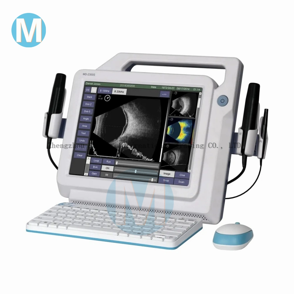
MD-2300S Ultrasonic A/B Scanner for Ophthalmology
Features
Specifications
Clinical Gallery
Vitreous Hemorrhage (10MHz)
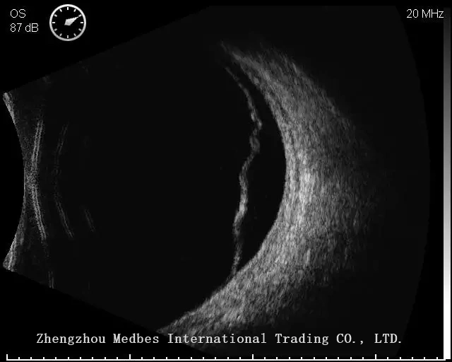
Vitreous Hemorrhage (20MHz)
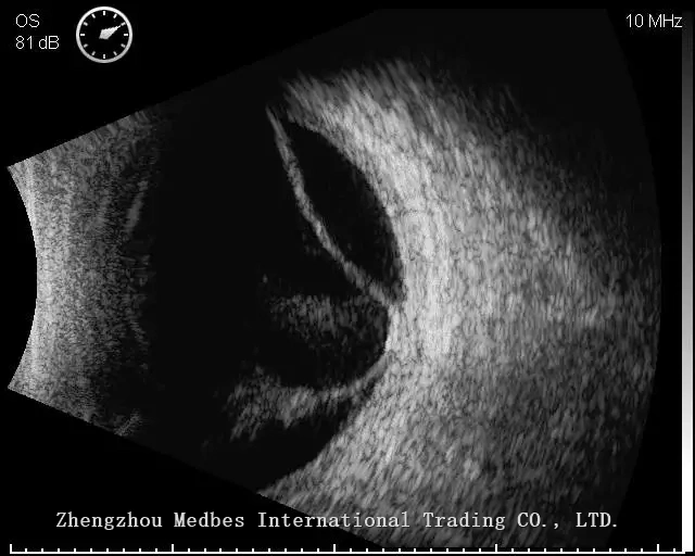
Posterior Vitreous Detachment (10MHz)
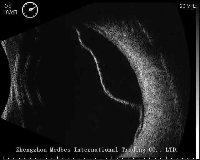
Posterior Vitreous Detachment (20MHz)
Retina Detachment (10MHz)
Retina Detachment (20MHz)
Certificate
CE, FDA, ISO13485 and CFDA
Options
Immersion Shell, Trolley, Video Printer, Graph/text Printer
Features
- High Definition 10MHz & 20MHz B-scan images
- High Resolution LCD Touch Screen
- Image/Video snapped and stored in real time in multiple buffer slots for immediate comparison and review
- Multiple TGC options for operator's preference including vitreous-enhanced mode
- Editable clinical report with A-scan/IOL results, B-scan images, and textual comments with configurable entries
- Reports in .PDF format for sharing and print-out
- Compatibility with graph/text printer
- Diverse connections via HDMI and USB 2.0 ports
- HDMI interface for double-screen showcase
- Massive storage capacity for over 20,000 exams
Specifications
| B-Scan | |
| Ultrasound Probes: | 10MHz B-Scan Probe New 20MHz B-Scan Probe |
| Detection Depth: | 10MHz:≥60mm;20MHz :≥20mm |
| Axial Resolution: | 10MHz:≤0.1mm;20MHz :≤0.08mm |
| Lateral Resolution: | 10MHz:≤0.2mm;20MHz :≤0.15mm |
| Scan depth: | 10MHz:34mm-60mm, 6-Level selectable 20MHz:22mm-40mm, 6-Level selectable |
| Scan Angle: | 53° |
| Cineloop & Image: | Capture and store up to 10s cineloop; Replay in real time, images displayed Continually or separately |
| Separately save individual frames from cineloop as an image | |
| Gray Scale: | 256 Levels |
| Gain: | 1-105dB adjustable |
| TGC: | Default, Vitreous-Enhanced Mode & Customized |
| AL Measurement: | 5-Point "Dict-5" Marking Method under B+A Mode |
| IOL Calculation: | SRK-II, SRK-T, BINK-II, HOLLADAY, HOFFER-Q, HAIGIS |
| Post-Refractive: | History-derived, Refraction-derived, SHAMMAS, Double K/SRK-T, ROSA |
| Post-processing: | Measurement (Angle, Area, Distance) Text Annotation & Image Magnification |
| Biometric A-Scan | |
| Ultrasound Probe: | 10MHz with Fixation Red Light |
| Gain: | 1-60dB |
| Measurement Method: | Contact or Immersion |
| Measurement Mode: | Manual or Automatic (Normal, Aphakic, Special and Cataract) |
| Measurements parameter: | Axial Length (AL), and Anterior Chamber Depth (AC), Lens Thickness (LEN), Vitreous Body Length (VITR), |
| Average and Standard Deviation for up to 10 scans per exam | |
| Configurable Tissue Velocities under Special or Manual mode | |
| AL Measuring Range: | 15mm-40mm |
| AL Measuring Accuracy: | ≤±0.05mm |
| General | |
| Display: | High-Resolution 12.1" LCD Touch Screen |
| Printer: | Graph/Text Printer and Video Printer (PAL) |
| Interface: | Video-Out (PAL), HDMI, USB2.0 Ports, DICOM 3.0 |
| Input: | Touch Screen, Footswitch, Wireless Mouse & Keyboard |
| HDD: | 320GB or higher |
| Power Supply: | AC 100-240V, 50/60Hz |
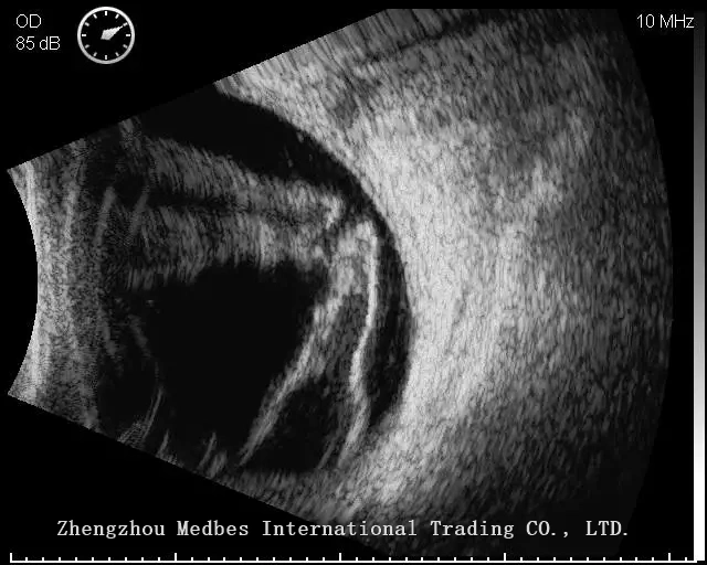
Clinical Gallery
Vitreous Hemorrhage (10MHz)
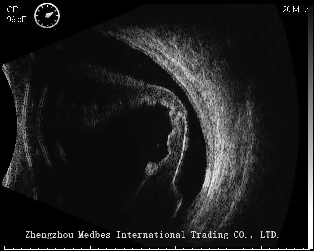
Vitreous Hemorrhage (20MHz)
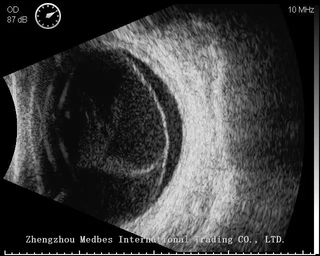
Posterior Vitreous Detachment (10MHz)
Posterior Vitreous Detachment (20MHz)
Retina Detachment (10MHz)
Retina Detachment (20MHz)
Certificate
CE, FDA, ISO13485 and CFDA
Options
Immersion Shell, Trolley, Video Printer, Graph/text Printer