Ophthalmic Equipment Optical Coherence Tomography With Efficient 3D Analysis
MC-OCT2000
Efficient automatic image acquisition
Easy guided macular or disc scan.
Quickly switch to anterior segment mode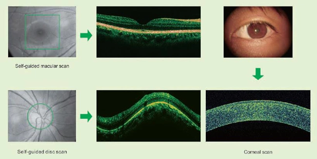
Precise positioning of the scan area, easy to focus on the lesion
Easily select the scan area for image acquisition.
Expanded field of view.
Focus on the lesion
Scan area positioning requires minimal
patients' cooperation
Double-click reset the scan area to the center.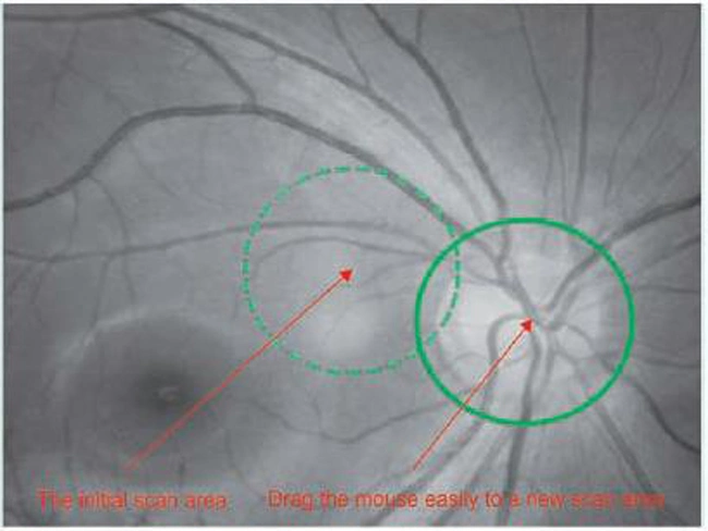
Plenty of report templates and editable modules
A variety of default report templates to expedite the report generation.
Template design feature facilitates the diagnoses writing.
Electronic reports in PDF format are convenient for doctors and patients to read and save.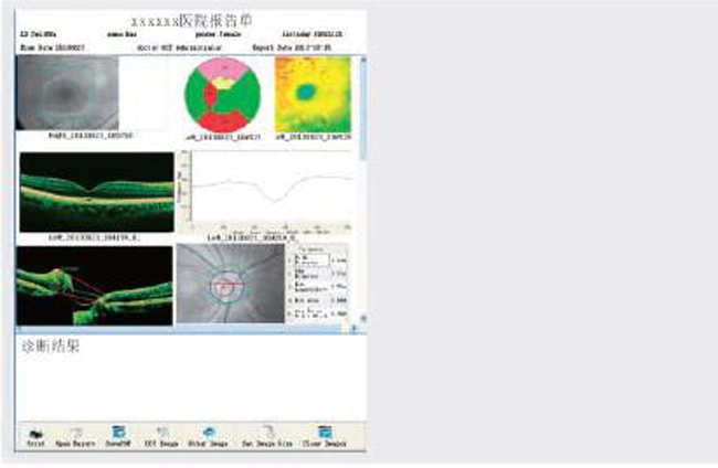
Unique community for consultation, teaching, and case study
Extensive cases to browse and search.
Enhanced teaching function.
Post and search comments to make consultation easier.
Unique lesion contour measurement function
You can quickly and easily correct the inaccurate contour lines generated by the automatic algorithm. The correct contour lines can be saved for future analysis, such as thickness topographic map and 3D map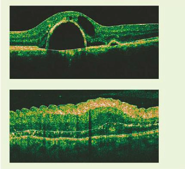
Unique disc edge identification function
Automatically measure the cup-disc ratio, rim area and other optometric parameters.
Automatically identify and draw the disc contour and cup contour in the fundus image.
You can correct the disc contour on the fundus map to accurately calculate the area of the disc and cup.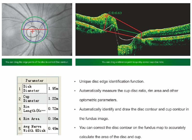
Precise follow-up scan technology
Automatically record the location of each inspection and repeat the data collection at the exact same locatior of the last inspection by using the image registration algorithm.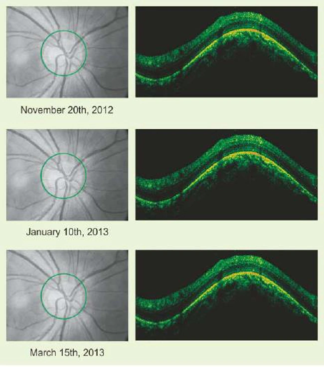
Anterior segment analysis
Angle measurement: You can measure the anterior chamber angle, distance between the iris and any point on posterior comeal surface, anterior chamber area.
Corneal measurement You can measure the corneal vertex thickness after refractive error correction and the thickness between any upper and lower surfaces.
Accurate Glaucoma analysis
A single measurement can automatically evaluate the average thickness of the retinal nerve fiber layer in each sector.
Glaucoma follow-up analysis and comparison between left and right eyes show intuitive diagnostic details.\
Efficient 3D analysis
3D analysis: Rotate, zoom, layered, sliced, reconstruction, retinal vascular map, save and print 3D map,3D topographic map report.
Specification of our OCT
| Resolution(in tissue) | Axial 5.0μm;Transverse:15μm |
| Scanning range | Depth 2.5mm;Wide≥6mm |
| Light source | SLD.840nm |
| Scanning speed | ≥27000 A-scans/sec. |
| Light power | 750μW on cornea |
| Fixation | Internal and external fixation |
| Minimun pupil diameter | 3mm |
| Scan pattern | 3D scanning,Radial scanning,Raster scanning.Circular scanning,HD scanning |
| Software analysis | Macular thickness topography, Average thickness of 9-sector, Macular thickness map, Disc thickness tomography, Disc thickness map, Circumpapillary clock hour distribution, Cup-to-disc ratio, Disc/cup/rim area, Comparative analysis of disc circular scan between left and right eyes, 3D analysis, Follow-up analysis, HD scan analysis. |
| Anterior segment module Scan pattern Software analysis | Cornea scanning, Anterior chamber angle scanning. Cornea thickness map, Cornea thickness topography, Cornea curvature topography, Cornea HD analysis, Anterior chamber angle analysis. |